Introduction
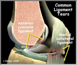
 The Collateral Ligaments are commonly injured structures in the knee. These injuries can occur in many ways. The injury usually involves a significant force, such as a fall while skiing or a direct force to the side of the leg.
Anatomy
Where are the collateral ligaments and what do they do?
Remember that the difference between a ligament and a tendon is that ligaments connect one bone to another bone while tendons connect muscles to bones. The collateral ligaments of the knee connect the femur bone on the top to the tibia bone on the bottom. The main purpose of these ligaments is to limit side to side motion of the knee. If these ligaments are stretched too far, they may tear. The tear may occur in the middle of the ligament, or it may occur where the collateral ligament attaches to the bone, on either end. If the force is great enough other ligaments may also be torn. The most common combination is a tear of the medial collateral ligament (MCL) and a tear of the anterior cruciate ligament (ACL).
The lateral collateral ligament (LCL) on the other side of the knee can also be torn, but it is less common than the medial collateral ligament. The ligament can be torn in other areas, similar to the medial collateral ligament.
Causes
How do collateral ligament injuries occur?
The collateral ligaments can be torn in sporting activities, such as skiing or football. This usually occurs when the lower leg is forced sideways – either towards the other knee (medially) or away from the other leg (laterally). A blow to the outside of the knee while the foot is planted can put stress on the medial collateral ligament (MCL) and result in a tear of the ligament. Slipping on the ice can cause the foot to slip outward, taking the lower leg with it. The body weight pushing down causes a force on the whole leg – just like bending a green stick. The medial collateral ligament (MCL) may be torn in this instance because the force hinges the knee open putting stress on the MCL.
Symptoms
What do Collateral Ligament Injuries feel like?
An injury violent enough to actually tear one of the collateral ligaments causes significant damage to the soft tissues around the knee. There is usually bleeding into the tissues around the knee, swelling of the tissues and perhaps bleeding into the knee joint itself. The knee is stiff and painful. As the initial stiffness and pain resides there may be a feeling of instability in the knee joint, and the knee may give away and not support your body weight.
Chronic (meaning long term, or persistent) instability due to an old injury to the collateral ligaments may also occur. If the torn ligament heals but is not tight enough do support the knee, a feeling of instability will persist. The knee will give away at times, and may be painful with heavy use.
Diagnosis
How do we look into this problem?
The initial physical examination usually gives a very good indication of which ligaments have been torn in and around the knee. In some cases, there is too much pain and muscle spasm to completely tell what is damaged in your knee. Your physician may suggest a period of rest with a knee splint and then re-examine the knee in 5 – 7 days. This will allow some of the initial pain and spasm to decrease and the exam may be more reliable. The swelling will also get better during this time allowing your doctor to better examine the knee.
X-rays may be required to rule out the possibility that bony damage has occurred as well. Stress X-rays may be beneficial to confirm that one of the collateral ligaments have been torn. Stress X-rays are simply plain x-rays taken with someone attempting to open the side of the joint that is suspected to be unstable. The X-rays will show a widening of the joint space on that side if instability is present.
An MRI scan may be ordered if there is evidence that multiple injuries have occurred, including injury to the meniscus or the anterior cruciate ligament. The MRI (Magnetic Resonance Imaging) machine uses magnetic waves rather than x-rays, to show the soft tissues of the body. With this machine, we are able to “slice” through the area we are interested in very clearly. Usually, this test is done to look for injuries, such as tears in the menisci or ligaments of the knee. This test does not require any needles or special dye, and is painless. If there is a uncertainty in the diagnosis following the history and physical examination, or if other injuries in addition to the collateral ligament tear are suspected, the MRI scan may be suggested.
Treatment
How do we treat Collateral Ligament Injuries?
Most injuries to the collateral ligaments will heal with simple immobilization in a cast or brace for 4-6 weeks. An isolated injury to the LCL or MCL infrequently requires surgical repair or reconstruction. The initial treatment for a collateral ligament injury focuses on decreasing inflammation in the knee. Rest and anti-inflammatory medications, such as aspirin, can help decrease the pain and swelling. As the ligament heals, a physical therapy program will help in decreasing pain and inflammation, improving motion, and regaining strength. Your brace may be locked so that you cannot bend the knee at first to help you avoid painful movements.
As your pain and swelling begins to go away a physical therapist may be contacted to help you with rehabilitation of the knee. Exercises to help regain normal movement of joints and muscles may be suggested at this point. When soreness is still present, these exercises must be done slowly and carefully to avoid further irritation. You will likely be instructed in a few home exercises to help with knee motion. As the soreness goes away, more vigorous stretching can be used to insure full movement in the knee.
The next part of your rehabilitation following injury will focus on strengthening the muscles around the knee. A set of exercises called closed kinetic chain exercises have become popular among physical therapists. These are exercises done with the foot fixed to the ground, while the body is moved in various directions to exercise the muscles. They generally do not require any fancy equipment and can be done at home. These exercises are designed to allow the muscles around the knee to be exercised while easing stress on the ligaments. These exercises are functional – because they represent activities we do throughout the day. Examples include stepping, squatting, lunging, and half kneeling.
The final stages of knee rehabilitation involve balance and proprioception exercises. Healthy ligaments send information to the brain about the position of a joint. This process is called proprioception. This partly explains how you know precisely where your finger is — even when your eyes are closed. Once a ligament has been injured, these nerves that do this are torn, and unable send the needed information to the brain. This increases the possibility of injury in the future. Balance and proprioception exercises help restore this position sense by retraining the nerves as they grow back. Examples of these types of exercises involve standing and walking on uneven or very soft surfaces, balancing on one leg, and jumping exercises.
If there are other injuries present, surgery may be required. Some orthopedists feel that if there is a combination of an ACL tear and an MCL tear, then both should be treated surgically. Others do not agree, and feel that the MCL tear should be treated with casting or bracing and the ACL reconstructed later. Time will tell if one approach is better than the other.
Repair of a freshly torn collateral ligament usually requires the surgeon to make an incision through the skin over the area where the tear in the ligament has occurred. If the ligament has been pulled from its attachment on the bone, the ligament is reattached to the bone with either large sutures or a special metal bone staple. Tears of the middle areas of the ligament are usually repaired by sewing the ends together.
Chronic instability caused by a collateral ligament injury may require a surgical reconstruction. A reconstruction differs from a repair of the ligaments. A reconstruction type operation usually works by either tightening up the loose ligaments or replacing the loose ligament with a tendon graft. If a tendon graft is needed, it is usually taken from somewhere else in the same knee. A very common graft that is used is the semitendinosis tendon. Studies have shown that this tendon can be removed without really affecting the strength of the leg. There are other, much bigger and stronger hamstring muscles that can take over the function of the tendon once it is removed. There are numerous different ways to perform a surgical reconstruction of either the lateral collateral ligament (LCL) or the medial collateral ligament (MCL) depending on which has torn.
After surgery for either repair or reconstruction of the collateral ligaments has been done, a physical therapist will help with your rehabilitation. As healing progresses, the rehabilitation program will be very similar to the program outlined earlier in this section for non-operative treatment.