Background
14 year old girl with acute injury to articular cartilage.
There are modern procedures that can in many cases restore areas of acutely injured articular cartilage in the knee. It should be noted that these are note meant for knees that have developed the secondary changes of osteoarthritis yet. Acute injuries to the normally smooth articular surface are known to lead to osteoarthritis eventually. Unfortunately, there is very poor healing potential in the hyaline cartilage of the knee articular surface once damage.
ACI
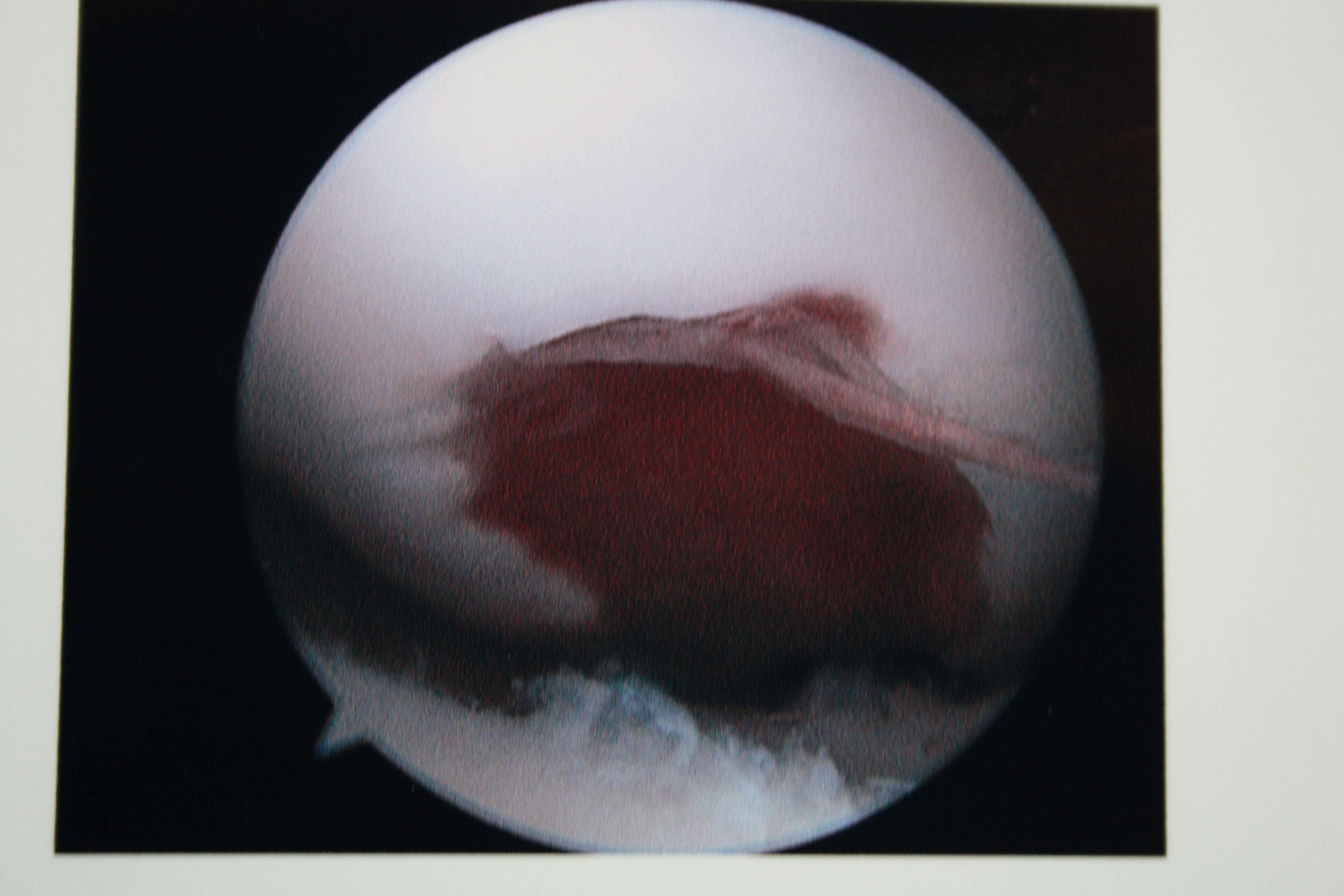
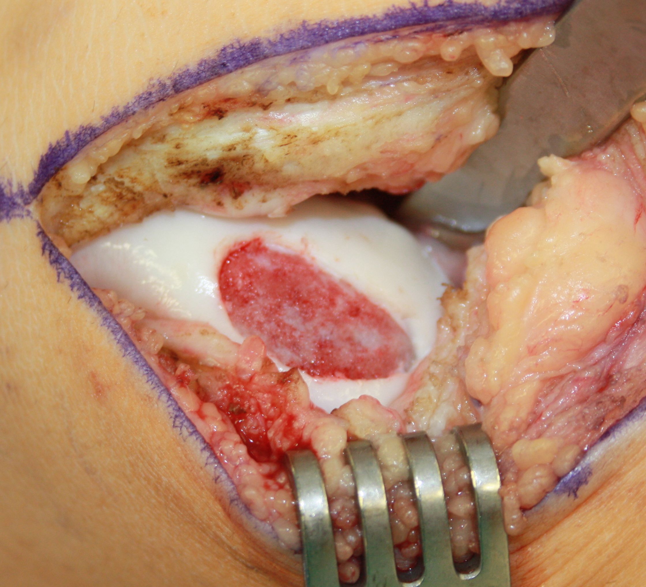
ACI or autologous chondrocyte implantation is one way to biologically restore more normal functioning acticular cartilage tissue. In the picture above, there is a full thickness, large osteochondral defect in this case of the lateral femoral condyle. You can see the defect hematoma that develops which will lead to the development of scar cartilage. This is the body’s normal attempt to heal this injury, but we know that scar cartilage doesn’t function like normal hyaline cartilage and will not last. ACI procedure will attempt to replace this scar cartilage with the patients own articular cartilage cells that have been grown in the lab. The following sequence of pictures will show steps of the procedure. Click image for larger view.
-
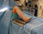
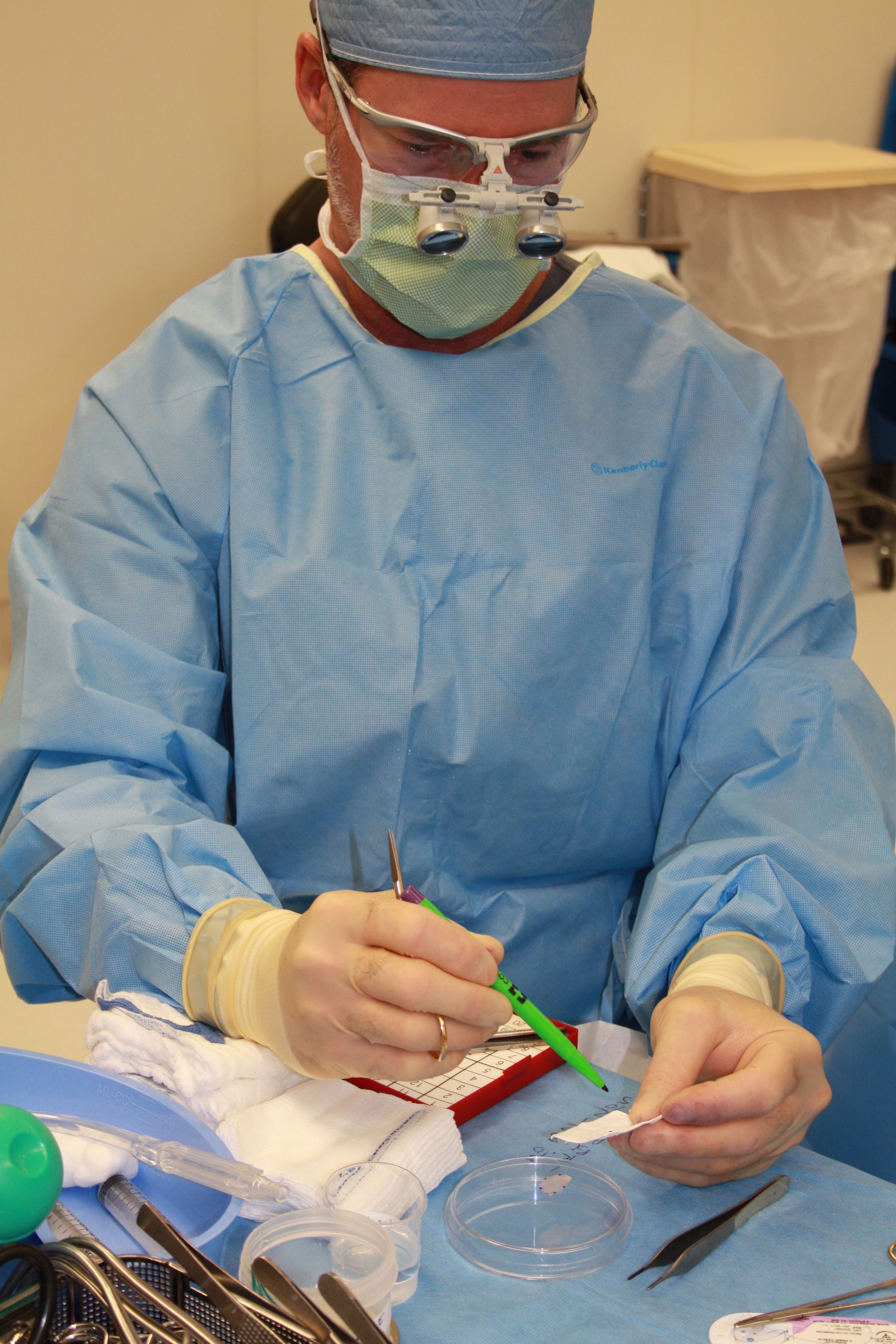
- Surgical set-up for ACI. Note the use of the Alvarado for easy change of flexion / extension

-

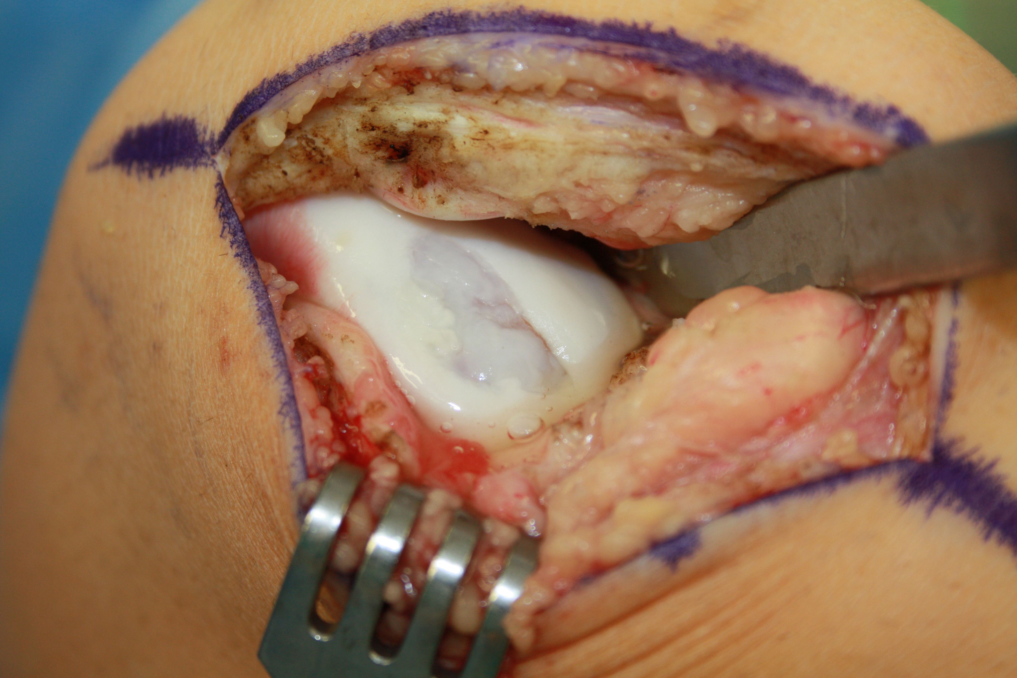
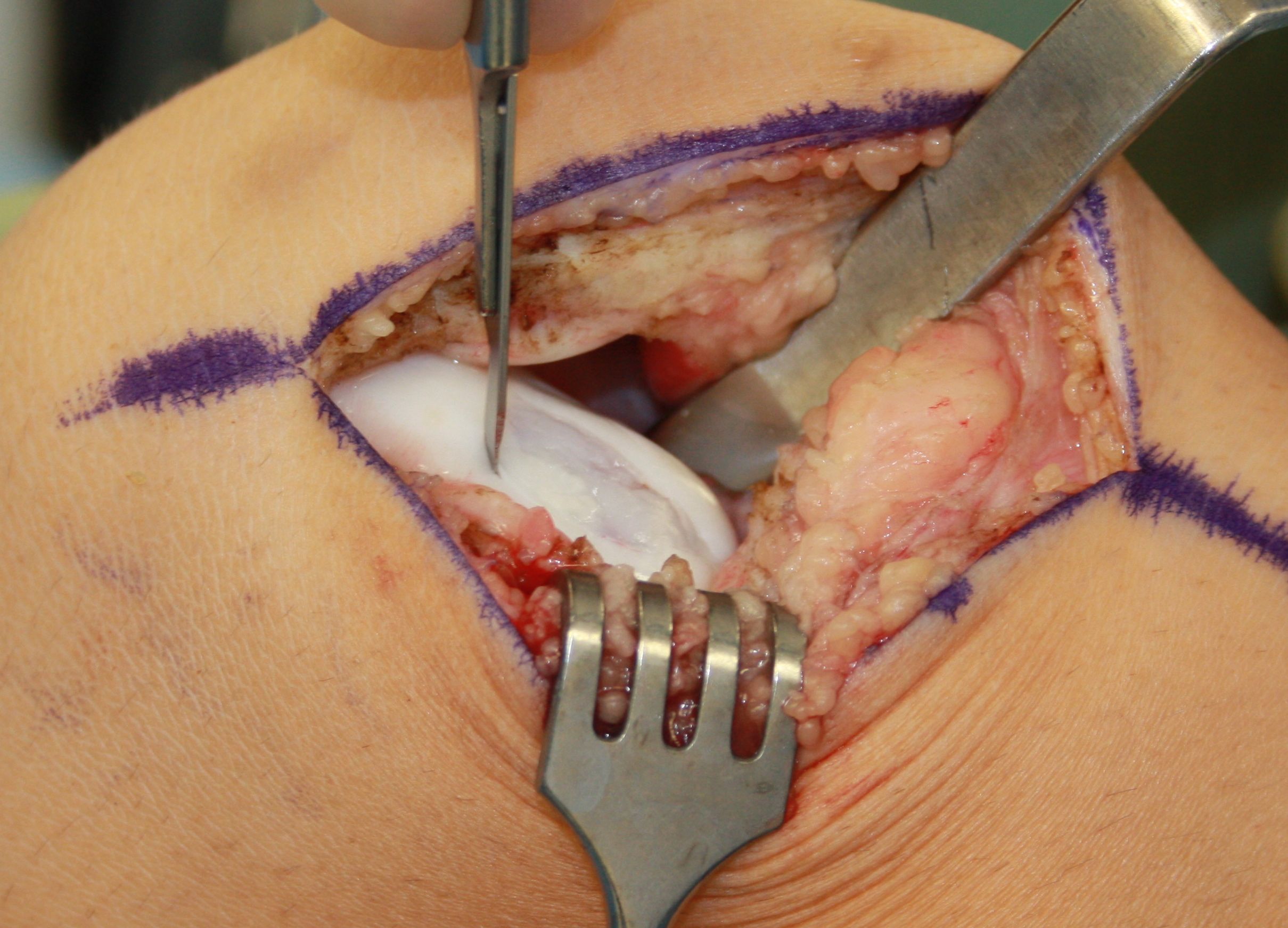
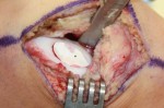
- Large osteochondral defect on the lateral femoral condyle of 14 yr old girl
-
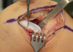
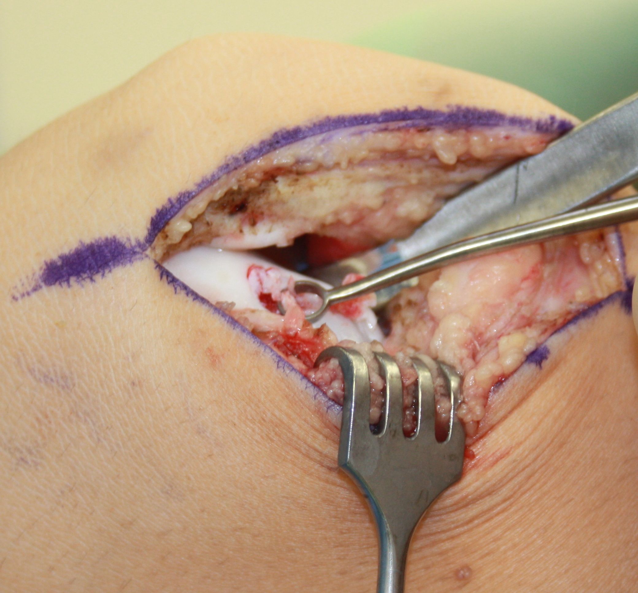
- Scalpel is used to delineate the defect to normal articular cartilage edges.
-

- A curette is used to remove all scar cartilage from defect including the calcified cartilage layer.
-
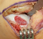
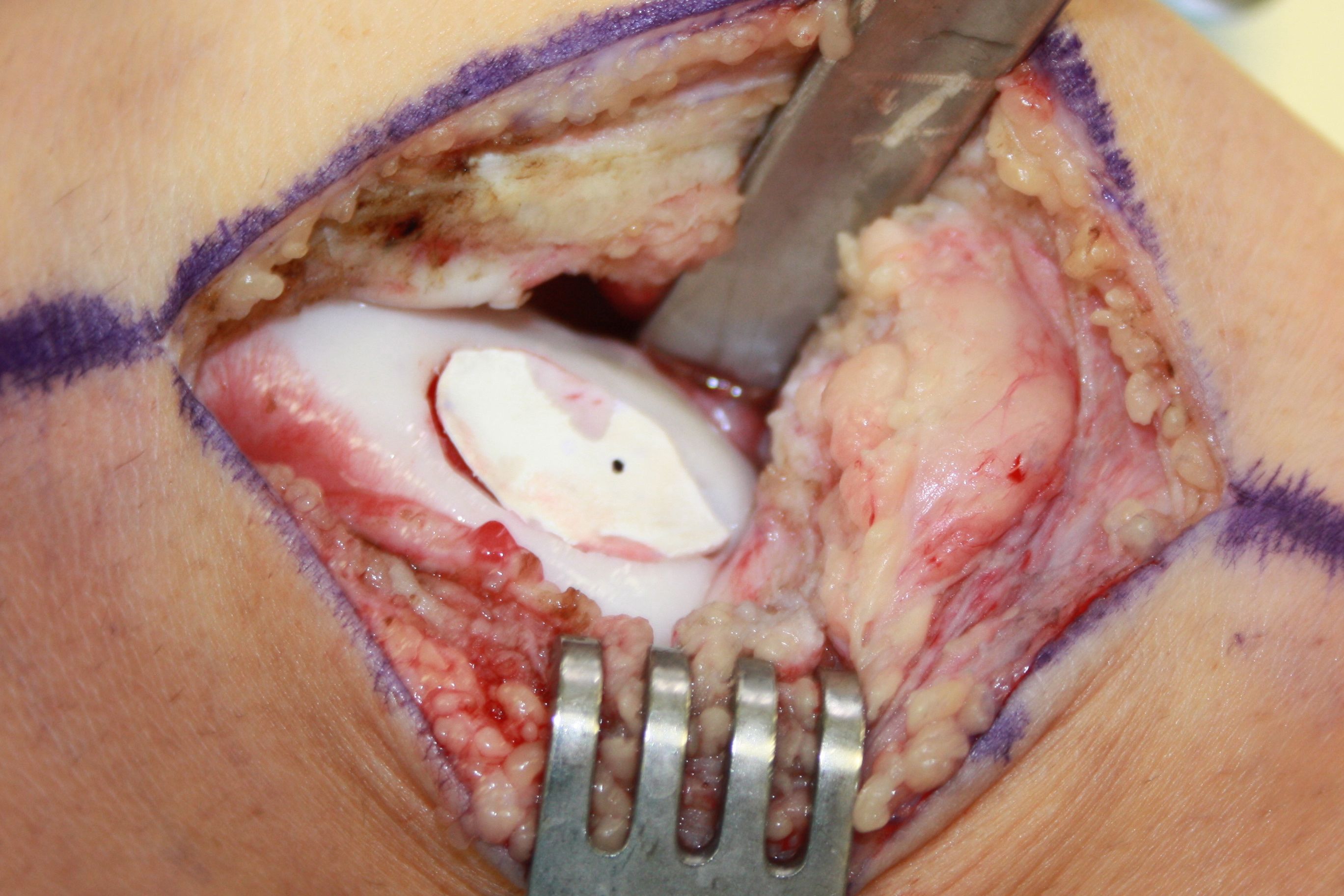
- Prepared defect ready for implantation.
-

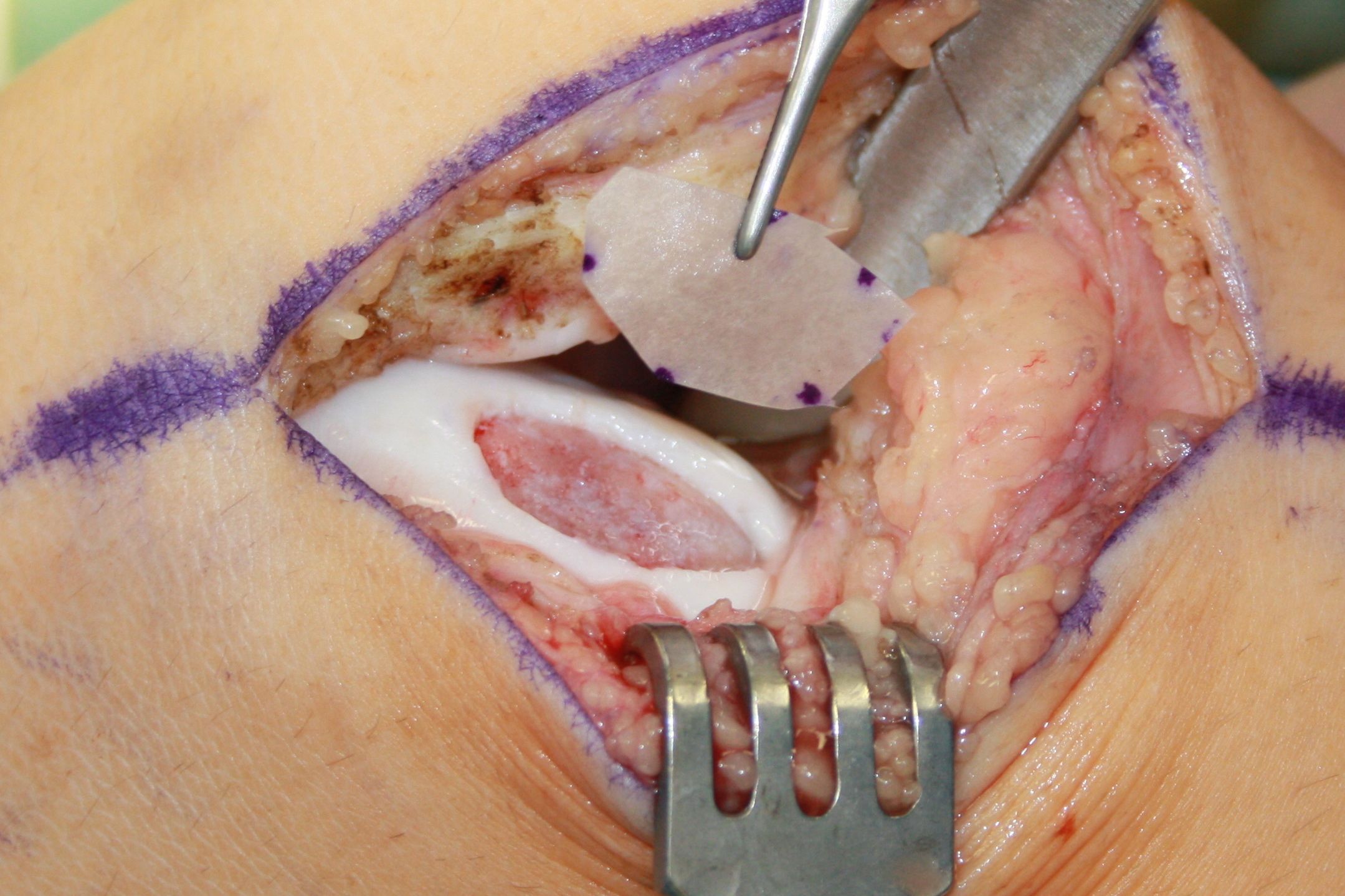
- Template is measured to size of defect.
-
- Dr. Maffet prepares biologic patch.
-
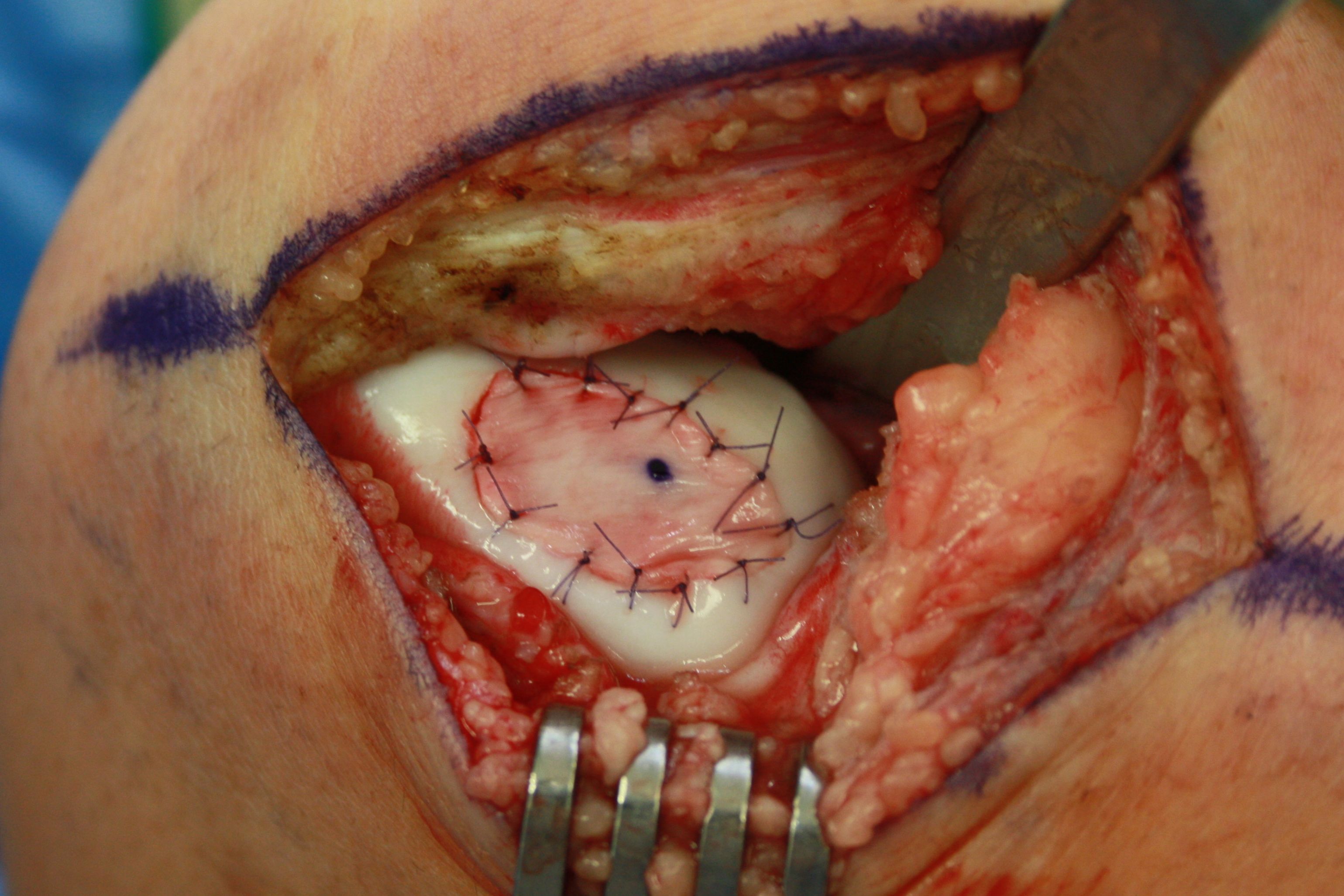
- Biologic patch in place over cartilage defect.
-

- Biologic patch is carefully sew in place creating a water tight seal from outside environment.
-
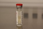
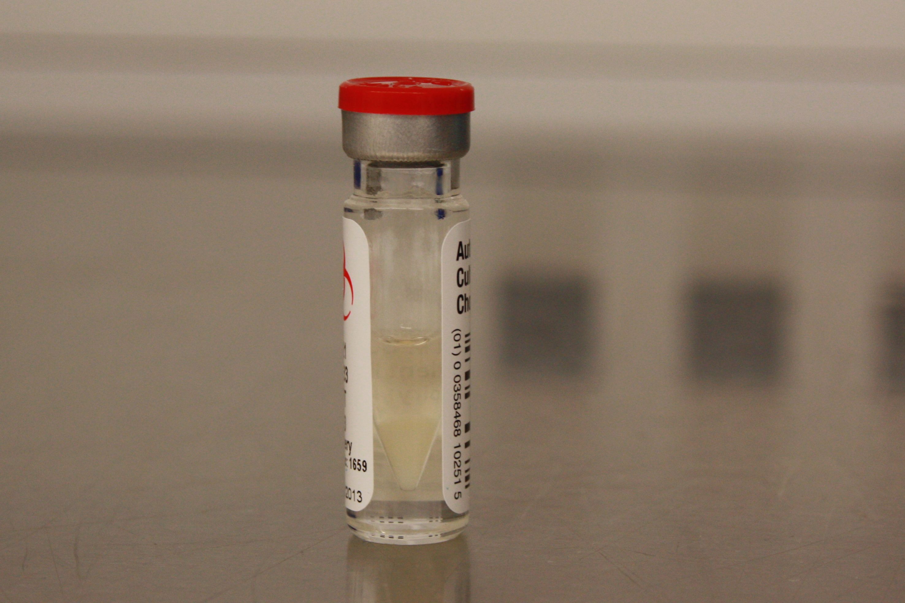
- Genzyme vial of millions of the patient’s hyaline cartilage cells which are introduced under the patch to grow on the prepared defect.